Myositis Ossificans
 A 46-year-old man reports that he experienced moderate pain of bilateral thighs when he rode his bicycle. The pain was associated with hard projections of the underlying muscles. Fifteen years earlier, he had sustained a pelvic fracture during a motor vehicle accident.
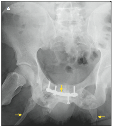
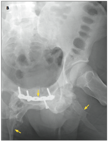
Physical examination revealed firm and mildly tender medial aspects of both thighs, which extended from the pubic symphysis to just above the knee joints. Radiographs showed heterotopic bone formation and calcification at the level of the pubic bone extending along the medial thighs and regions of the sartorius musculature (A and B). Myositis ossificans was subsequently diagnosed.
Myositis ossificans traumatica is a non-neoplastic proliferation of bone and cartilage in an area previously exposed to trauma and/or hematoma. It primarily
affects the proximal limb muscles. Myositis ossificans traumatica is believed to result from passive stretching.1
This condition is encountered most frequently in adolescent and young adult males because of a sports-related injury. It can also be found in various other
patient populations as a result of often forgotten trauma to muscle tissue. Sometimes, it may be mistaken for a serious disease, such as sarcoma.
The frequency of the development of myositis has not been well documented; however, after a direct blow to a muscle, the reported incidence is 9% to 17%.1 The incidence also is thought to be related to the severity of injury.
 Clinically, myositis should be suspected if the pain and swelling after a traumatic injury do not respond to conservative treatment within a week or so of initial trauma that is limited to the muscles. Radiographic abnormalities can be seen within 18 to 21 days of injury, and definite radiographic evidence should be present by 2 months. The pathologic features of myositis ossificans traumatica also have been described.1
Treatment of myositis ossificans traumatica largely is based on the RICE principle of rest, ice, compression, and elevation. Excision maybe indicated if the mass is large enough to predispose the affected area to additional injury or if it limits adjacent joint range of movement to the extent that it could cause functional disability. It is universally accepted that surgical excision should not be done until the ossification of the muscle or bone mass is mature. Maturity is confirmed by diminished activity in a bone scan, which usually takes about 6 to 12 months.2 Removal of immature bone often results in extensive local recurrence. This patient decided not to undergo an orthopedic evaluation for surgical excision.
1. Beiner J, Jokl P. Muscle contusion injury and myositis ossificans traumatica. Clin Orthop Relat Res. 2002;(403 suppl):S110-S119.
2. Tyler JL, Derbekyan V, Lisbona R. Early diagnosis of myositis ossificans with T099m diphosphonate imaging. Clin Nucl Med. 1984;9:460-462.


