Peer Reviewed
Innumerable Septic Pulmonary Emboli
AUTHORS:
Zachary Field, MD, and Mario Madruga, MD
Department of Internal Medicine, Orlando Regional Medical Center, Orlando, Florida
CITATION:
Field Z, Madruga M. Innumerable septic pulmonary emboli [published online February 11, 2020]. Consultant360.
A 39-year-old woman presented to the emergency department with generalized weakness, fevers, and a productive cough over the past 3 days. Her medical history was significant for intravenous drug use, methicillin-resistant Staphylococcus aureus (MRSA) endocarditis, and hepatitis C virus infection.
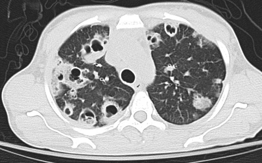
A computed tomography scan of the thorax showed innumerable cavitary lesions throughout the lungs (Figure) consistent with multiple septic pulmonary emboli (SPE). She was found to be septic, with blood cultures growing MRSA.

Figure. A thoracic computed tomography scan showed innumerable cavitary lesions throughout the patient’s lungs, consistent with multiple SPE.
The patient was started on intravenous vancomycin and cefepime. Echocardiography was performed, which showed a normal ejection fraction and a large, pedunculated, mobile mass on the anterior and septal tricuspid valve leaflet consistent with endocarditis. The patient underwent tricuspid valve replacement. She was evaluated by an infectious disease specialist, who switched antibiotics to ceftaroline, which the patient continued for 6 weeks.
Discussion. SPE is a rare disease process in which microorganisms in a fibrin matrix are mobilized from an infected site throughout the vasculature and imbed in the pulmonary vascular network.1 SPE has a high mortality rate of approximately 10%.2 Several processes have been associated with SPE, including intravenous drug use, translocation of periodontal bacteria, device-related infections, infective endocarditis, and skin and soft tissue infections.2,3 These emboli can lead to localized pulmonary abscess formation and lung infarction.3
The most common microorganisms associated with SPE are S aureus, coagulase-negative Staphylococcus species, Streptococcus viridans, Candida species, Fusobacterium species, and Klebsiella pneumoniae.2,3 Treatment includes broad-spectrum antibiotics, generally with a combination of penicillins and cephalosporins, for a period of 4 to 9 weeks.1,2 For blood cultures that are positive for MRSA, systemic intravenous vancomycin or daptomycin is recommended.2
REFERENCES:
- Mattar CS, Keith RL, Byrd RP Jr, Roy TM. Septic pulmonary emboli due to periodontal disease. Respir Med. 2006;100(8):1470-1474. doi:10.1016/j.rmed.2005.1010
- Ye R, Zhao L, Wang C, Wu X, Yan H. Clinical characteristics of septic pulmonary embolism in adults: a systematic review. Respir Med. 2014;108(1):1-8. doi:10.1016/j.rmed.2013.10.012
- Ojeda Gómez JSA, Carrillo Bayona JA, Morales Cifuentes LC. Septic pulmonary embolism secondary to Klebsiella pneumoniae epididymitis: case report and literature review. Case Rep Radiol. 2019;2019:5395090. doi:10.1155/2019/5395090


