Myositis Ossificans Traumatica
A previously healthy 8-year-old boy presented after having injured his left arm while playing football 2 weeks prior. Radiographs obtained at that time showed no fractures. Erythema, swelling, and warmth had developed in the arm over the several days after the injury, but he had not developed fever, chills, nausea, diarrhea, or other systemic symptoms.
The patient had a history of an infection “on the side of the knee” approximately 2 months previously, which had been drained in the office of his primary care pediatrician and had been treated with antibiotics.
Physical examination revealed a nondiscrete soft-tissue mass, approximately 2 × 4 cm in size, that was deep to muscle and tender on palpation. Grip strength in the left hand was normal; left biceps and triceps strength were rated as 3/5. Range of motion at the elbow was limited due to pain, and the patient was unable to lift his left arm over his head.
Laboratory test results at admission included a white blood cell count of 7,100/µL. The erythrocyte sedimentation rate was elevated at 32 mm/h, and the C-reactive protein level was 4.6 mg/L. He received an initial diagnosis of bacterial infection, given the swelling, erythema, and warmth over the affected area and the history of an abscess 2 months prior. He was treated empirically for Staphylococcus aureus infection with intravenous clindamycin along with an ibuprofen suspension for pain.
The following morning, plain radiographs showed considerable soft tissue swelling at the medial midshaft region of the left humerus (A). There was a faint curvilinear calcification within the soft-tissue swelling (arrow). There was no cortical erosion, destruction, or trabecular distortion at the site. Calcifications within the hematoma were thought to be the most likely cause of the symptoms, but soft tissue tumor could not be excluded.

The presenting radiograph, taken 2 weeks after injury, showing soft-tissue swelling at the medial midshaft region of the left humerus. A faint curvilinear calcification is visible within the soft tissue (arrow). (Image courtesy of Jonathan Williams, MD)
Magnetic resonance imaging done the same day showed the mass to be poorly defined and completely within the belly of the triceps muscle, with peripheral enhancement and marked perilesional edema extending distally and proximally from the mass. A diagnosis of myositis ossificans traumatica (MOT) was made.
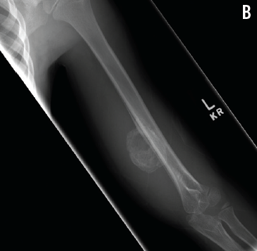
The patient’s pain and range of motion both improved with the administration of ibuprofen, and he was referred to an orthopedic specialist for outpatient follow-up. Over the following 3 weeks, his symptoms abated, and radiographs were consistent with maturing myositis ossificans (B). At 3 months’ follow-up, the mass had begun to shrink, and at 2 years from the initial presentation, it had completely resorbed.

Radiograph of the humerus, taken 6 weeks after injury, denoting maturing myositis ossificans with increased peripheral mineralization and progressive periosteal reaction. (Image courtesy of Charles Bush, MD)
Myositis ossificans (MO) is characterized by heterotopic bone formation via non-neoplastic processes and most commonly is caused by trauma.1 MO is rare in children and occurs most frequently in young athletes who play contact sports.2 MOT is the most common form of MO, representing 60% to 75% of cases.3
The differential diagnosis is broad, and MOT often is misdiagnosed initially. The most common misdiagnosis in published case studies has been osteosarcoma, followed by osteomyelitis, soft-tissue sarcoma, cellulitis, and lymphadenitis.2 Radiographs give the appearance of a soft-tissue mass, with faint calcifications appearing up to 6 weeks after the onset of symptoms.4 As the lesion matures, immature fibrous tissue develops at the center, with the periphery consisting of mature-appearing bone. Eventually, most of the heterotopic bone resorbs by 5 to 6 months after the onset of MOT without intervention.5
MOT is best treated conservatively with nonsteroidal anti-inflammatory drugs to reduce pain and inflammation. Referral to an orthopedic specialist may be necessary. n
References
1. Gindele A, Schwamborn D, Tsironis K, Benz-Bohm G. Myositis ossificans traumatica in young children: report of three cases and review of the literature. Pediatr Radiol. 2000;30(7):451-459.
2. Micheli A, Trapani S, Brizzi I, Campanacci D, Resti M, de Martino M. Myositis ossificans circumscripta: a paediatric case and review of the literature. Eur J Pediatr. 2009;168(5):523-529.
3. Goldman AB. Myositis ossificans circumscripta: a benign lesion with a malignant differential diagnosis. AJR Am J Roentgenol. 1976;126(1):32-40.
4. Ackerman LV. Extra-osseous localized non-neoplastic bone and cartilage formation (so-called myositis ossificans): clinical and pathological confusion with malignant neoplasms. J Bone Joint Surg Am. 1958;40(2):279-298.
5. Chadha M, Agarwal A. Myositis ossificans traumatica of the hand. Can J Surg. 2007;50(6):E21-E22.


